Pigmentation patterns of shells of mollusks - a natural picture book to study dynamic systems

A special case of biological pattern formation is the emergence of the pigment patterns on the shells of molluscs. These patterns are of great diversity and frequently of great beauty. The shells consist of calcified material. The animals can increase the size of their shells only by accretion of new material along a marginal zone, the growing edge of the shell. In most species, pigment becomes incorporated during growth at the edge. In these case, the pattern formation proceeds in a strictly linear manner. The second dimension is a protocol of what happens as function of time along the growing edge. The shell pattern is, so to say, a space-time plot. The shells provide a unique situation in that the complete history of a highly dynamic process is preserved. This provides the opportunity to decode this process.
In normal development, a strong evolutionary pressure exists to reproduce faithfully a given structure. In contrast, the functional significance of the pigment patterns on shells is not clear. There is presumably no strong selective pressure to preserve a given shell pattern. Thus, nature was able to play. Although the patterns look overtly very different, it is to be expected that similar molecular mechanism are at work. The challenge was to find corresponding models. With some additions to the standard patterning reactions, the models were able to describe many patterns in great detail (Meinhardt and Klingler, 1987 ). [PDF] . An extensive treatment including software is given in a book (Meinhardt, 2003).
ELEMENTARY PATTERN ON THE SEA SHELLS: TRACES OF A STABLE PATTERN, OSCILLATIONS AND TRAVELING WAVES
Basic elements of the shell patterns are lines parallel, perpendicular or oblique to the direction of growth. Keeping in mind their space-time character, lines parallel to the direction of growth (and thus usually perpendicular to the growing edge, left figure below) indicate that pigment production occurs at particular positions that are separated by regions without pigment production. This is the usual situation as discussed above for normal development. In contrast, pigmented lines perpendicular to the direction of growth (usually parallel to the growing edge, central figure) are traces of a more or less synchronous oscillation in pigment production. In terms of the model, this occurs if the antagonistic reaction follows the self-enhancing reaction too slowly. The activation increases in an avalanche-like manner. It is only somewhat later that the accumulating inhibitor (or the removal of all the substrate) causes a collapse in the activator production. A refractory phase follows. A new activation can be triggered only after the decay of the remaining inhibitor. In other words, a longer time constant of the antagonistic reaction can lead to oscillations. A rapid diffusion of either the activator or the inhibitor can cause their synchronization.

Lines oblique to the growing edge result from traveling waves of pigment production (right figure). In the model, they result if the activator has a small diffusion range while the antagonistic substance is nearly non-diffusive. An activated region can "infect" its neighboring region such that, after a certain lag phase, it becomes fully activated too, and so on. The situation is very similar to the wave-like spread of an epidemic. One person can infect its neighbors. The full development of a sickness is also based on a self-enhancing effect, the replication of the virus. Some time after bursting virus proliferation, the immune system begins to acts antagonistically. It captures the virus and the person will become healthy again. For the spread of the epidemic it is crucial that only the virus, but not the immune response is transmitted from one individual to the next. In the time record on the shells, this leads to oblique lines. V-like tips are the record of an annihilation of two waves.
A common aspect in many of these patterns is the involvement of a second antagonistic reaction that extinguishes concentration maxima shortly after they have been build up, causing the permanent modification of the spatial pattern. The following shell is an example:

In the simulation above it is assumed pattern formation takes place by an activator-depletion mechanism. In addition, a local but long lasting inhibitor poisons newly-formed maxima. A homogeneous oscillation (centre) becomes unstable in favor of an out-of-phase oscillation. A somewhat higher substrate production leads to a transition from the out-of-phase oscillations to travelling waves (right). The same transition is visible on the shell (left). The two antagonistic effects, depletion and inhibition, lead to the superposition of a pattern in time and a pattern in space. It is easy to see that in both the checkerboard pattern and in the oblique line pattern (travelling wave) an alternation of activated and non-activated regions occurs along the space and along the time coordinate. The modeling of these patterns has turned out to be a key for the understanding of other pattern forming processes, for instance, phyllotaxis and the orientation of chemotactic cells and growth cones.
On many species, the pattern documents an unusual behavior. Two examples will be discussed below.
BRANCHING LINES: SUDDEN TRIGGER OF BACKWARDS-RUNNING WAVES


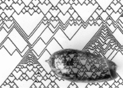
The shell of Oliva porphyria document sudden trigger of a backwards-running waves. Usually two colliding waves annihilate each other (V-like pattern elements) since waves cannot enter regions that are refractory due to the counter wave. The splitting of waves is a strategy to maintain the number of traveling waves. In the model, a global measure of how many waves are present can be obtained from a hormone that is produced by all activated cells. If the number of waves and thus this concentration drops below a critical value, the system shifts from an oscillating into a steady state modus (by a feedback on the half life of the inhibitor). Activated regions remain activated until a backwards-running waves are triggered. This leads to a doubling of the number of waves and thus to a return in the oscillating mode. The model describes that many branches are formed simultaneously even at distant positions. The simulation above shows the situation in the pigment-producing mantle gland. The activator-inhibitor system (black-red) forms traveling waves that extinguish each other. If too few waves are left, the hormone concentration (green) becomes very low. This causes that cells remain activated until the backwards-running wave is initiated. For a simple BASIC program in which this algorithm is used to generates this pattern click here
CROSSING OF LINES: WAVES THAT PENETRATE EACH OTHER DURING COLLISION


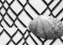
The shell of Tapes literatus is decorated with oblique lines that cross each other. Again, this is unusual since, as mentioned, waves in excitable media usually annihilate each other upon a collision. A simulation of this behavior is possible under the assumption that two antagonists are involved. In addition to the fast and long ranging inhibition, a second antagonist is assumed. It causes locally a de-activation of an activation. The activation can disappear at this and reappear at a displaced position. (This situation will be discussed again in connection with phyllotaxis and the orientation of chemotactic cells and growth cones ). Alternatively, the activation can be shifted into a neighboring region that is not yet poised. In the latter case, traveling waves result that appear normal. However, if two waves collide, the situation is different. The decline of the activator (black) causes a rapid drop in the long ranging inhibition (red). This causes the survival of the activation during collision until two new diverging waves are triggered. In this simulation, the local antagonistic reaction results from the depletion of a necessary component (green). In the time record, crossing lines result. The model accounts also for a global perturbation seen on the specimen shown: many lines terminate at a particular growth line while others bifurcate at same instance. Such a behavior occurs after a sudden general reduction of the activation since this reduces also the long-range inhibition (for details and software, see Meinhardt, 2003 ).
Further Reading and References
Meinhardt, H. and Klingler, M. (1987). A model for pattern formation on the shells of molluscs. J. theor. Biol 126, 63-69. [PDF]
Meinhardt, H. (2003). The Algorithmic Beauty of Sea Shells. 3rd enlarged edition Springer, Heidelberg, New York. (with PC - software)
An interesting website describing many shell patterns has been provided Philippe Guyot-Sionnest:
http://oliva.porphyria.free.fr/; also links to other website can be found there.


