Midline formation in Planarians: a circumventing DV border has organizing functions
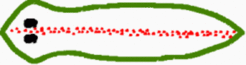
Not much information is available on midline formation in planarians. One of the relevant findings is that a BMP homologue has been cloned in Dugesia japonica that forms a sharp midline on the dorsal side along most of the AP axis (drawing after Orii et al. [1]):

Planarians are well known for their almost unlimited capability for regeneration [2,3]. Most remarkable, the overall hierarchy of the AP and DV axes seems to be completely different from that observed in insects and vertebrates. In planarians a confrontation between dorsal and ventral cells is the precondition that anterior- or posterior-most terminal regions regenerate [4,5] while in vertebrates the ancestral AP pattern can be formed without any DV pattern [6]. The available data suggest that in planarians the lateral positions are determined first and midline formation is a secondary event. The figure below illustrates the proposed mechanism (Meinhardt, 2004):


A primary DV subdivision (pink/blue) is assumed. The resulting DV border (green) obtains organizing properties. It is the precondition to initiate the most anterior (A, blue) and most posterior (P, brown) structures. Intercalation of the pattern along the AP axis leads to the long extension of the animal. The dorsal midline (red) is localized by an inhibitory influence from the circumventing organizing border (green). In the simulation below a stripe-forming system (red) is assumed. The stripe emerges at the largest possible distance from the circumventing DV border. It sharpens in the course of time. If the terminal signals (blue/brown) weekens the repulsive effect, the midline can extend to the most anterior and posterior positions.

Further Reading and References
Meinhardt, H. (2004). Different strategies for midline formation in bilaterians. Nat Rev Neurosci 5, 502-510 [PDF]
- Orii, H., Kato, K., Agata, K. & Watanabe, K. (1998). Molecular cloning of bone morphogenetic protein (BMP) gene from the planarian Dugesia japonica.[2] Baguñá, J., Salo, E., Romero, R., Garcia-Fernandez, J., Bueno, D., Muñoz-Marmol, A.M., Bayas-Casramirez, J.R. & Casali, A. (1994). Regeneration and pattern-formation in planarians - cells, molecules and genes. Zool Sci 11, 781-795.
- Agata, K., Tanaka, T., Kobayashi, C., Kato, K. & Saitoh, Y. (2003). Intercalary regeneration in planarians. Dev. Dyn. 226, 308-316.
- Chandebois, R. (1979). The dynamics of wound closure and its role in the programming of planarian regeneration. Develop. Growth and Differ. 21, 195-204.
- Kato, K., Orii, H., Watanabe, K. & Agata, K. (2001). Dorsal and ventral positional cues required for the onset of planarian regeneration may reside in differentiated cells. Dev. Biol. 233, 109-121.
- Ober, E.A. & Schulte-Merker, S. (1999). Signals from the yolk cell induce mesoderm, neuroectoderm, the trunk organizer, and the notochord in zebrafish. Dev. Biol. 215, 167-181.