p9.htm

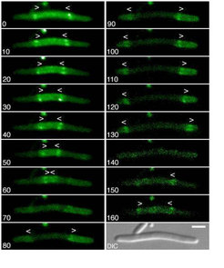
Time-lapse fluorescence micrographs showing the dynamic behavior of MinE-Gfp in a short filamentous cell. Times are indicated in seconds. Note the accumulation of MinE-Gfp in two dynamic E-rings as well as in an extra-annular peripheral pattern (PEA signal) which is present either in between two rings (0-60s, and 150-160s), or in between a ring and the proximal cell pole (80-130s). Note the net movement of rings towards the PEA signal(s) (0-60s, 80-130s, and 150-160s), the disappearance of rings when they either approach each other (60s), or one of the cell poles (130s), and the subsequent appearance of new rings and PEA signal(s) in the region(s) previously devoid of signal (80 and 150s). Arrowheads (< or >) indicate both the position of a ring as well as the direction of its movement.
Shown is a filament of strain DR102/pDB346/pDR174 [DminCDE::aph, ftsZ0, recA::Tn10 /PlR::ftsZ, cI857/Plac ::bfp-minD, minE-gfp]. Cells were grown in the presence of 50mM IPTG at 30oC and displayed a filamentous phenotype due to the depletion of FtsZ. The bar in the DIC panel represents 2mm. For additional details, see [1]
References
- Hale, C. A., Meinhardt, H., and de Boer, P. A. J. (2001). Dynamic localization cycle of the cell division regulator MinE in E. coli., EMBO J. 20, 1563-1572.